Foot anatomy
|
|
|
(Biel, A, 2010 P. 354)
|
http://www.britannica.com/EBchecked/media/101314/Bones-of-the-foot-showing-the-calcaneus-talus-and-other
|
Bones
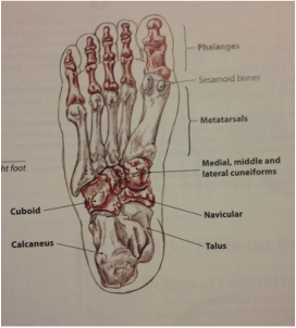
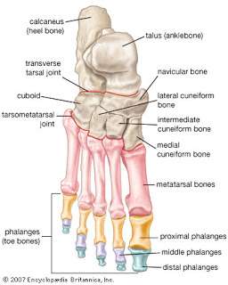
Toes: The toes are formed by separate bones called phalanges. The first toe, or hallux, contains only proximal and distal phalanges, while the four other toes are made up of three phalanges, proximal, middle and distal.
Metatarsals: These five bones (1st-5th) consist of a proximal base, a shaft and a distal head. The metatarsals articulate with the phalanges and the tarsal bones and make up a majority of the forefoot. There are also two small sesamoid bones located on the dorsal side of the head of the 1st metatarsal.
Tarsals: The calcaneus, talus, navicular, cuboid, and medial, middle, and lateral cuneiforms are known as the tarsal bones of the foot, which are located between the metatarsals and lower leg bones.
- Calcaneus: large chunky bone that forms the heel of the foot.
- Talus: Sits superior to the calcaneus and articulates with the tibia and fibula of the lower leg forming the talocrural, or ankle joint.
- Navicular: bean-shaped bone located between the medial and middle cuneiforms and the talus.
- Cuboid: cube-shaped bone is surrounded by the 4th and 5th metatarsals, lateral cuneiform and the calcaneus.
- Cuneiforms: The three cuneiforms lie in a row between the navicular and metatarsals and make up the "Archway" of the foot.
Muscles
The intrinsic muscles of the foot allow for toe flexion, and extension and foot inversion, eversion, abduction, adduction, supination, and pronation.
(Starkey et al, 2010)
(Starkey et al, 2010)
(Starkey et al, 2010, P. 134-138)
http://toplowridersites.com/related-pictures-of-a-skeletal-muscle-cell-the-cell-membrane-is-called/
http://www.hpssandiego.com/ligaments_and_muscles_of_the.htm
http://gophoto.us/key/dorsal%20foot%20anatomy
http://toplowridersites.com/related-pictures-of-a-skeletal-muscle-cell-the-cell-membrane-is-called/
http://www.hpssandiego.com/ligaments_and_muscles_of_the.htm
http://gophoto.us/key/dorsal%20foot%20anatomy
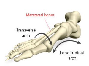
Arches
There are both longitudinal and transverse arches of the foot. The arches are supported and maintained by the shape of the foot bones, the muscles of the foot, and numerous ligaments. An individual with little to no arches, or "flat feet", may have a subtle arch which appears normal when sitting then becomes flat upon standing, or a rigid arch which always appears flat. Having low or fallen arches can create pain from lack of support in the foot (see injuries).
There are both longitudinal and transverse arches of the foot. The arches are supported and maintained by the shape of the foot bones, the muscles of the foot, and numerous ligaments. An individual with little to no arches, or "flat feet", may have a subtle arch which appears normal when sitting then becomes flat upon standing, or a rigid arch which always appears flat. Having low or fallen arches can create pain from lack of support in the foot (see injuries).
The lateral longitudinal arch is located on the lateral side of the food. It is typically very flat and does not contain many bones.
The transverse arch is not a true arch but is formed by the three cuneiforms, the cuboid, and the five metatarsal bases. This arch is supported by the interosseous, dorsal, and plantar ligaments.
The transverse arch is not a true arch but is formed by the three cuneiforms, the cuboid, and the five metatarsal bases. This arch is supported by the interosseous, dorsal, and plantar ligaments.
|
http://sportspodiatryinfo.wordpress.com/2010/08/09/the-transverse-metatarsal-arch/
|
http://sportspodiatryinfo.wordpress.com/2010/08/09/the-transverse-metatarsal-arch/
|
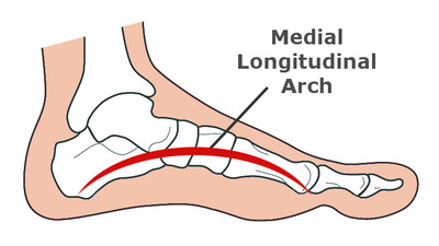
The medial longitudinal arch is on the medial side of the foot and is very tall. It is supported by the plantar fascia, and various plantar ligaments. The medial longitudinal arch may be very subtle on some and excessively high on others. This arch serves to support the foot and act a spring to aid in walking and running.
Articulations
Subtalar Joint: Synovial joint which allows only three degrees of movement, supported by the lateral and medial talocalcanean ligaments.
Midtarsal Joints: Consist of the Talocalcaneonavicular, cuneonavicular, cuboideonavicular and the intercuneiform, cuneocuboid, and calcaneocuboid joints.These joints allow for gliding and rotation to give the foot its accessory motion.
Tarsometatarsal Joints: Plane synovial joints which are gliding joints. Also referred to as Lisfranc's joint.
Intermetatarsal Joints: Composed of four plane synovial joints allow for gliding
Metatarsophalangeal Joints: Composed of five condyloid synovial joints. The movements at these joints are flexion, extention, abduction, and adduction.
Interphalangeal Joints: Synovial hinge joints, there is one IP joint on the hallux and two, a proximal interphalangeal and a distal interphalangeal, on each of toes 2-5.
(Magee, 2008)
Other Structures
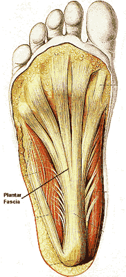
Plantar Aponeurosis: A thick superficial band of fascia stretching from the heel to the ball of the foot. Originates from the tuberosity of the calcaneus and expands toward the metatarsal heads and supports the longitudinal arch of the foot. Also known as the plantar fascia, this structure stabilizes the arch and flexes the first metatarsal allowing for it to carry a majority of the body weight and also acts as a shock absorber when the foot hits the ground.
Blood Supply: The posterior and anterior tibial artery, the peroneal artery, popliteal artery, plantar artery and the dorsal pedis artery supply the blood to the foot.
(Magee, 2008)
https://www.southerncross.co.nz/AboutTheGroup/HealthResources/MedicalLibrary/tabid/178/vw/1/ItemID/129/Plantar-fasciitis-heel-spur-syndrome.aspx
