Ankle & Lower Leg anatomy
Bones
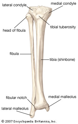
Tibia: The medial, larger bone of the lower leg. The proximal portion of the tibia is tibial plateau which acts as a cusp for the knee, the distal portion tapers into the medial malleoli and the concave surface which articulates with the talus at the ankle joint.
Fibula: The smaller lateral bone of the lower leg. The head of the fibula serves as an attachment point for the lateral collateral ligament of the knee and a number of muscles, the distal portion is known as the lateral malleoli.
Talus: The most superior of the tarsal bones, the talar dome articulates with the concave surface of the distal tibia to form the talocrural or ankle joint.
Calcaneus: The heel of the foot is a large chunky bone which forms an inferior articulation with the talus. The calcaneus serves as an attachment point for the Achilles tendon and takes bears most of the body weight.
http://anatomy.wikispaces.com/Tibia+and+Fibula
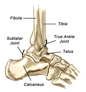
Bony structure of the lateral ankle
http://www.kidport.com/reflib/science/humanbody/skeletalsystem/Ankle.htm |
Anterior view of the lower leg, ankle, and foot
(Biel, A. 2010, P. 346) |
Muscles
The muscles of the ankle and lower leg allow for ankle dorsiflexion, plantarflexion, eversion, and inversion
The muscles of the ankle and lower leg allow for ankle dorsiflexion, plantarflexion, eversion, and inversion
https://myhealth.alberta.ca/health/pages/conditions.aspx?hwid=tp13087
(Starkey et al, 2010, P. 176-177)
Articulations
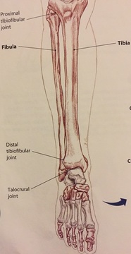
Tibiofibular Joint: Superior and inferior tibiofibular joints are where the tibia and the fibula articulate. Both are syndesmosis joints and are supported by the anterior and posterior tibiofibular ligaments and the inferior transverse and interosseous ligaments. There is little movement at the inferior tibiofibular joint but it allows for a small spread of the bones at the ankle joint during dorsiflexion.
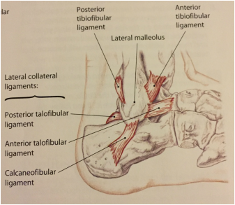
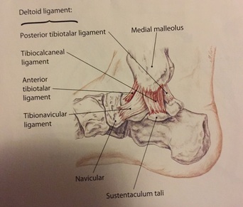
Talocrural (Ankle) Joint: Uniaxial, modified hinge, synovial joint located between the talus, medial malleolus of the tibia and the lateral malleolus of the fibula. This joint allows for dorsiflexion and planterflexion of the ankle. The ankle joint is supported by numerous ligaments:
Subtalar Joint: A synovial joint between the talus and the calcaneus which allows for inversion and eversion of the ankle
(Magee, 2008, p. 845-846)
Tibiofibular Joint: Superior and inferior tibiofibular joints are where the tibia and the fibula articulate. Both are syndesmosis joints and are supported by the anterior and posterior tibiofibular ligaments and the inferior transverse and interosseous ligaments. There is little movement at the inferior tibiofibular joint but it allows for a small spread of the bones at the ankle joint during dorsiflexion.
Talocrural (Ankle) Joint: Uniaxial, modified hinge, synovial joint located between the talus, medial malleolus of the tibia and the lateral malleolus of the fibula. This joint allows for dorsiflexion and planterflexion of the ankle. The ankle joint is supported by numerous ligaments:
- The medial ankle ligament is the deltoid ligament which is composed of four separate ligaments: the tibionavicular, tibiocalcanean, posterior tibiotalar, and anterior tibiotalar
- The lateral ankle ligaments are the anterior talofibular, posterior talofibular, and the calcaneofibular
Subtalar Joint: A synovial joint between the talus and the calcaneus which allows for inversion and eversion of the ankle
(Magee, 2008, p. 845-846)
(Biel, A. 2010, P. 398)
|
(Biel, A. 2010, P. 398)
|
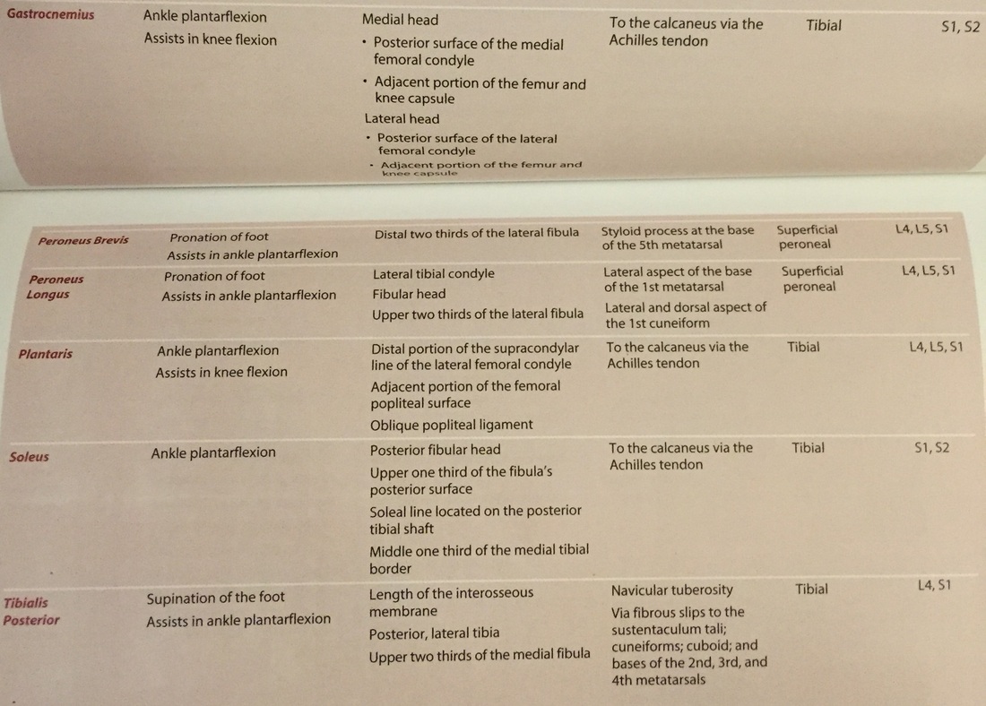
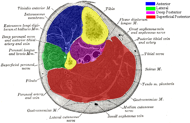
Compartments of Lower Leg
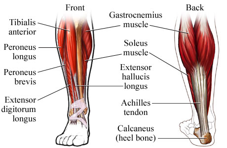
Anterior Comparment: muscles include the tibialis anterior, extensor hallucis longis, extensor digitorum longus, and peroneus tertius. The anterior compartment also houses the deep peroneal nerve and the anterior tibial blood vessels.
Lateral Compartment: Structures include the peroneus longus brevis muscles and the superficial peroneal nerve.
Superficial Posterior Compartment: Muscles include the gastrocnemius, plantaris, and soleus. This compartment also holds the sural nerve.
Deep Posterior Compartment: Muscles include the tibialis posterior, flexor hallucis longus, flexor digitorum longus, and popliteus. Also include in the deep posterior compartment is the tibial nerve and the poterior tibial vessels.
(Prentice, 2011)
http://php.med.unsw.edu.au/medwiki/index.php?title=File:Compartments_of_the_Leg.png
Nerve and Blood Supply
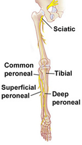
The nerve innervation of the lower leg comes via the common peroneal nerve which branches into the superficial and deep peroneal nerves.
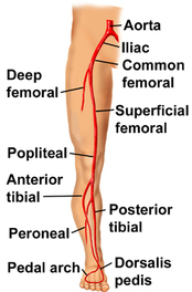
The ankle and lower leg blood supply comes via the anterior tibial artery and the posterior tibial artery.
The nerve innervation of the lower leg comes via the common peroneal nerve which branches into the superficial and deep peroneal nerves.
The ankle and lower leg blood supply comes via the anterior tibial artery and the posterior tibial artery.
|
|
http://www.yoursurgery.com/Procedures/lower_bypass/images/LegArteries.jpg
http://www.yoursurgery.com/procedures/longbonefracture/images/LegNerves.jpg
http://www.yoursurgery.com/procedures/longbonefracture/images/LegNerves.jpg
